


Osteoarthritis of the hip joint (ATS) is a slow and destructive disease. Under the influence of a number of reasons, in the course of the development of the disease, irreversible changes occur in the structure and properties of hyaline cartilage, which lead to an increase in pressure on the articular surfaces and their deformation or fusion. Given that mechanical overload is considered one of the main causes of the development of the disease, the joint of the hip joint is often affected by arthrosis.
Features of the anatomical structure of the hip joint
The hip joint (TC) is the junction of the pelvis and femur. This joint allows you to reduce and widen the lower limbs, lift the legs and bring them closer to the body and perform walking movements. From birth and throughout life, a person bears a large load on the hip joint.
From the side of the pelvic bone, the "acetabular" cavity takes part in the joint, from the side of the femur - its epiphysis. The acetabulum has a lip of collagen along its edges, which acts as a sort of seal that holds the femoral epiphysis tightly in its indentation. The notch in the center of the acetabulum is covered by a collagen membrane and is the attachment site of the ligament of the femur.
The composition of the TS capsule includes ligaments:
- femoral-iliac - the strongest ligament that can withstand a load of more than 200 kg and prevents excessive arching of the hip back;
- femoral-pubic - is responsible for the abduction and reduction of the thigh, thus limiting its circular movements;
- ischial femoral - protects the vehicle from concussions, reduces the load when walking and running;
- circular (loop) - prevents dislocations and holds the femoral head in the cavity of the pelvic cavity and is the basis of the joint bag.
Numerous muscle groups and tendons allow the vehicle to move in three axes:
- Longitudinal (vertical).
- Transverse (horizontal, frontal).
- Sagittal (anterior-posterior).
Joint arthrosis can occur both in a healthy joint and become a continuation of existing diseases of the musculoskeletal system.
What is this disease?
Hyaline cartilage performs cushioning and protective functions against damage to the joint surfaces. ATS is a disease in the process of development of which changes the structure of collagen cartilage fibers, which subsequently leads to their fragmentation and destruction. Fragments of cartilaginous fibers, if they get into the joint cavity, can cause an inflammatory process. Bare surfaces experience changes in bone tissue caused by friction and increased pressure. The cartilaginous tissue that remains along the edges of the epiphyses grows compensatory with subsequent ossification, causing ankylosis (immobilization of the bony junction). In the later stages, in the absence of adequate therapy, the patient completely loses mobility and becomes disabled. Destructive processes are provoked by various reasons.
There are the following types of arthrosis of the hip joint:
- Primary. Its etiology is not fully understood. Idiopathic (primary) osteoarthritis develops in a previously healthy joint. Most often, it develops in older people.
- Secondary. It is provoked by previous diseases of the articular apparatus, congenital anomalies of development, changes in the work of organs and systems of human vital activity.
The disease develops in one joint or affects both at the same time.
Causes of the disease
Among the causes contributing to the onset of the disease and its progression, the following are identified:
- Hereditary genetic predisposition to the development of the disease.
- Injuries of the bone joint (dislocations, fractures, sprains and tendons).
- Unbearable systematic potency and physical activity.
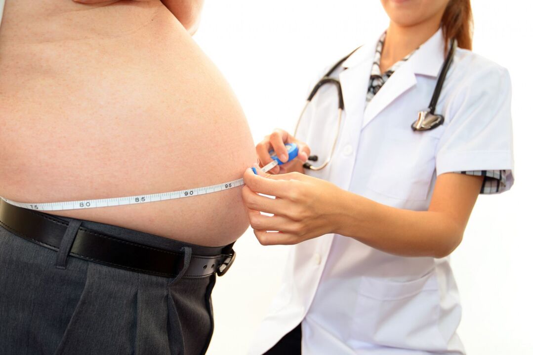
- Overweight.
- Functional disorders of the endocrine system (diabetes, psoriasis).
- Congenital pathologies of the structure and development of the musculoskeletal skeleton.
- Professional characteristics of labor activity.
- Poor local circulation.
- Previous diseases caused by pathogenic flora.
- Legg-Calve-Perthes disease.
- Metabolic disorders (gout).
- Physical inactivity.
- Immune diseases.
These reasons are not always able to cause ATS. Most often, the activation of pathological processes can be provoked by:
- increased stress and physical activity;
- constant overwork;
- hypothermia of the vehicle or the body as a whole;
- sudden lifting of heavy objects;
- hormonal imbalance;
- radiation exposure.
Symptoms of the disease
The symptomatic manifestations of ATS are similar to the manifestations of osteoarthritis of other joints.
The main characteristic symptoms of this disease are considered:
- Stiffness in the morning or after a long period of immobility.
- Decreased range of motion, change in gait.
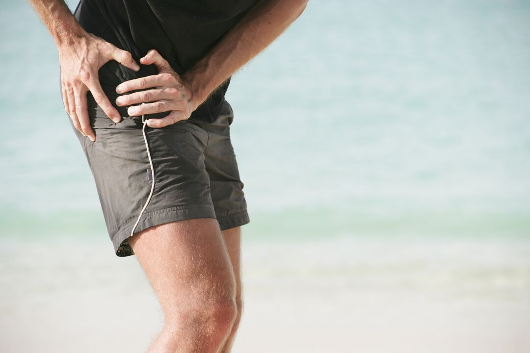
- Pain, first caused by mechanical or physical stress, then constant.
- The manifestation of creaks, crunches and clicking sounds during sudden movements.
- Pronounced lameness on the affected limb.
- The occurrence of contractures (limitation of passive movements).
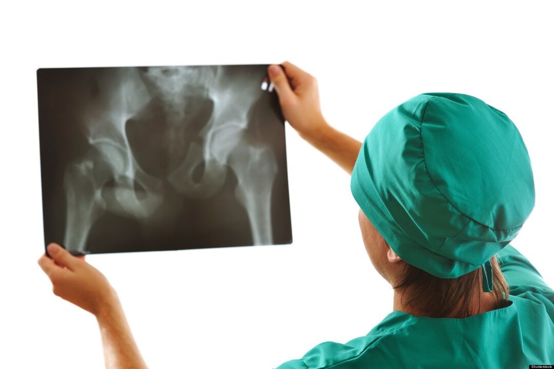
- Narrowing or closure of the joint space (radiographic sign).
The severity of the signs of arthrosis of the hip joint depends on the degree of development of the disease and the reactive capabilities of the patient's body.
Stages of coxarthrosis
Depending on the clinical manifestations, 4 stages of arthrosis of the hip joint can be distinguished:
- Arthrosis of the 1st degree of the hip joint does not have pronounced pain and other manifestations. The stage is difficult to diagnose, the disease can be detected by a biochemical study of hyaline cartilage tissue and the determination of an insufficient amount of glycosaminoglycans. The patient feels pain in the joint and rarely pain at the beginning of physical activity.
- Arthrosis of the second degree of the hip joint is characterized by changes in the density and elasticity of the cartilaginous fibers. Cracks and breaks appear. The depreciation functions are reduced. The pain intensifies, radiates to the inguinal region, the dilution and reduction movements of the affected limb are limited.
- At the third degree, the stratification of cartilaginous fibers occurs with greater intensity. The articular surfaces experience excessive pressure, foci of ischemia develop. Cartilaginous tissue grows along the edge of the epiphyses. The sensation of pain in the area of \u200b\u200bthe damaged bone junction does not depend on the state of activity and rest. With each movement, the joint "creaks" and "creaks". Movement range is reduced in all axes.
- The fourth degree is characterized by the exposure of the surfaces of the articular components with the formation of ulcers and depressions. The articular head of the femur is poorly fixed in the acetabulum, which leads to a violation of the confrontation and separation of the articular surfaces. During this period, the patient experiences excruciating pain caused by narrowing, sometimes closing of the joint lumen and compression of nerve fiber bundles and blood vessels. Movement is restricted, sometimes completely.
Classification of pathological changes caused by ATS is necessary to understand the mechanism and features of the development of the disease. Determining the severity of the disease helps determine the correct tactics of treatment and disability (in case of severe disease).
Possible consequences
The progression of ATS leads not only to the deformation of the femoral head and the pelvic cavity, but also to the development of pathological processes in the functioning of the articular apparatus as a whole.
Pathologies resulting from complications of hip arthrosis:
- synovitis (inflammation of the synovial membrane of the joint);
- aseptic necrosis of the femoral head;
- joint destruction (osteonecrosis);
- inflammation of the joint bag with changes in the amount of synovial fluid;
- partial or complete ankylosis (immobilization of the bone joint);
- contractures (limited mobility and inability to flex-extend the limb).
The development of complications of ATS always leads to a deterioration in the general condition of the patient, his quality of life and loss of movement without assistance.
Diagnostic methods
Diagnosis of osteoarthritis of the hip joint in the initial stage is difficult. Symptomatic manifestations become noticeable only when the epiphyses of bones and nerve fibers are involved in the pathological process.
During a medical examination in the progression phase, the following are found:
- visual change in joint contour;
- pain on palpation;
- sometimes softness of the periarticular tissues;
- shortening of the diseased limb.
The main role in the diagnosis of ATS is assigned to the X-ray examination. As auxiliary diagnostic methods used:
- Ultrasound, MRI.
- CT.
- Puncture of joint lubrication (synovial fluid).
- Diagnosis by arthroscope (microprobe).
- Clinical and biochemical laboratory tests of urine, blood.
Timely diagnosis improves the prognosis of treatment and the further life of the patient.
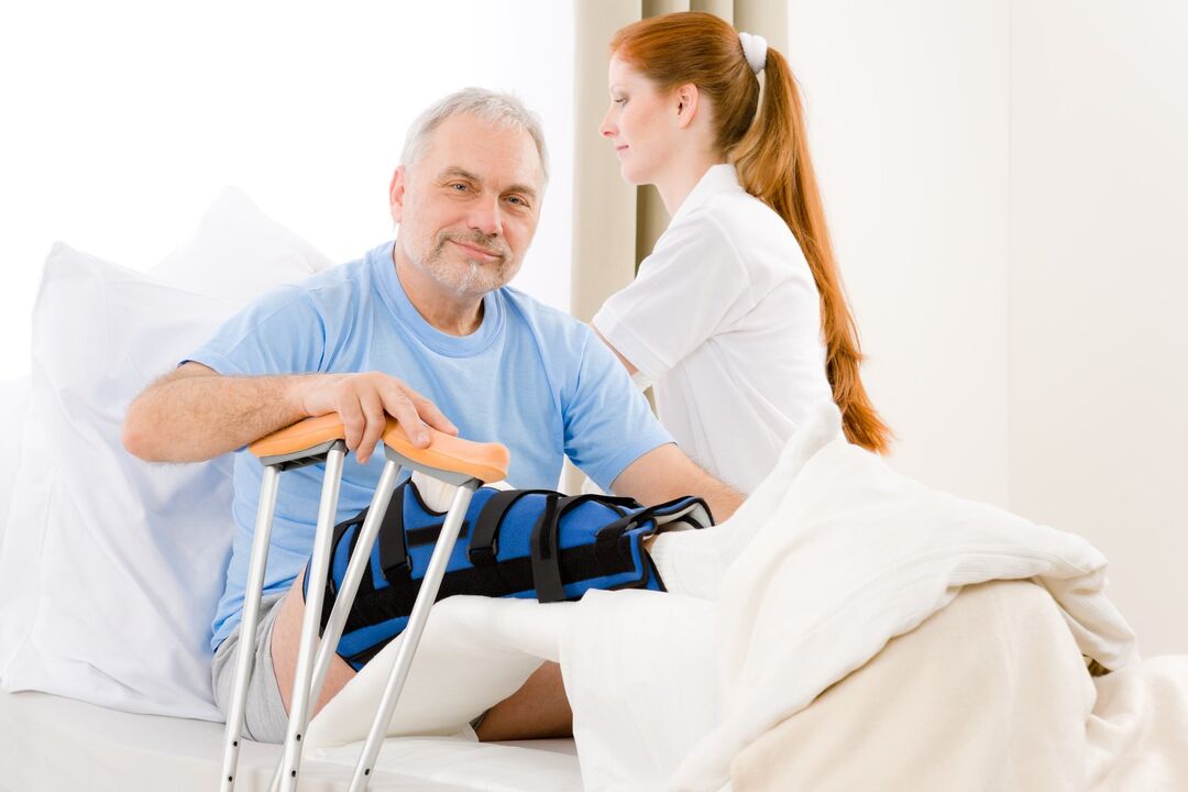
How to apply for disability?
It is impossible to cure this disease completely. To confirm the right to social benefits and assign a disability group after passing the examination by narrow specialists, you should contact your doctor.

The indication for the assignment of disability in case of arthrosis of the hip joint is:
- oligoarthrosis (injury of no more than 2 joints) TS 2 degrees;
- combined arthrosis of the 2nd degree of the ST and 3rd degree arthrosis of the knee joint;
- a decrease in the length of the diseased limb by more than 6 cm;
- responsive, documented sliding automatic telephone switchboard.
In determining the disability group will help:
- carefully collected medical history;
- the conclusion of the Medical Advisory Commission (MCC);
- results of diagnostic studies;
- passing the Medical and Social Expert Commission (MSEC).
If the decision of the commission of experts is negative, it can be appealed to the higher authorities.
Prevention
Preventive measures are an easy way to avoid the development of this disease. Preventive measures include:
- Adherence to an active lifestyle.
- Control of indicators of body weight.
- Optimization of nutrition and methods of work and rest.
- Reduced mechanical and physical load.
- Treatment of diseases of viral and infectious etiology.
- Prevention and prevention of injuries at home and at work.
- Regular preventive examination.
Conclusion
The answer to the frequently asked question: "Is it possible to cure arthrosis of the hip joint? " Experts give a negative answer. Destroyed cartilage tissue cannot be fully restored, just as it is impossible to completely correct the deformation and destruction of the bones included in the joint. Do not ignore even the minor manifestations of hip arthrosis, this reduces the chances of preventing further development of the disease.